
Introduction: When Artificial Intelligence Looks Deeper Than the Human Eye
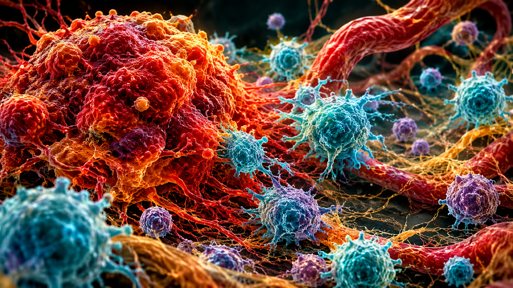
AI cancer detection is transforming medical research by helping scientists identify abnormal cellular patterns in microscopic tissue samples.
For decades, pathologists have relied on their experience, specialized staining techniques, and microscopic examination to identify cancer. Although this approach remains central to cancer diagnosis, interpreting tissue samples can be challenging. Tumors are often heterogeneous, meaning that cancerous and non-cancerous cells may exist side by side, while subtle changes in cellular structure can make some abnormalities difficult to identify.
Artificial intelligence (AI) is introducing a new approach to this process.
Using deep learning, computer vision, and advanced image-analysis techniques, AI systems can examine digitized tissue samples at extremely high resolution. They can identify patterns in cell shape, tissue architecture, tumor boundaries, and interactions between different cell populations.
The objective is not simply to make computers recognize cancer. Researchers are investigating whether AI can extract biological information from tissue images that may be difficult to quantify consistently through conventional microscopic examination.
This emerging field, known as computational pathology, is creating new possibilities for cancer detection, classification, prognosis, and personalized treatment.
But how does AI actually recognize cancer at the cellular level? Can it identify microscopic abnormalities that even experienced pathologists might overlook? And how close are these technologies to becoming routine clinical tools?
1. Understanding Cancer at the Cellular Level
To understand how AI detects cancer, it is important to first understand what pathologists look for when examining tissue.
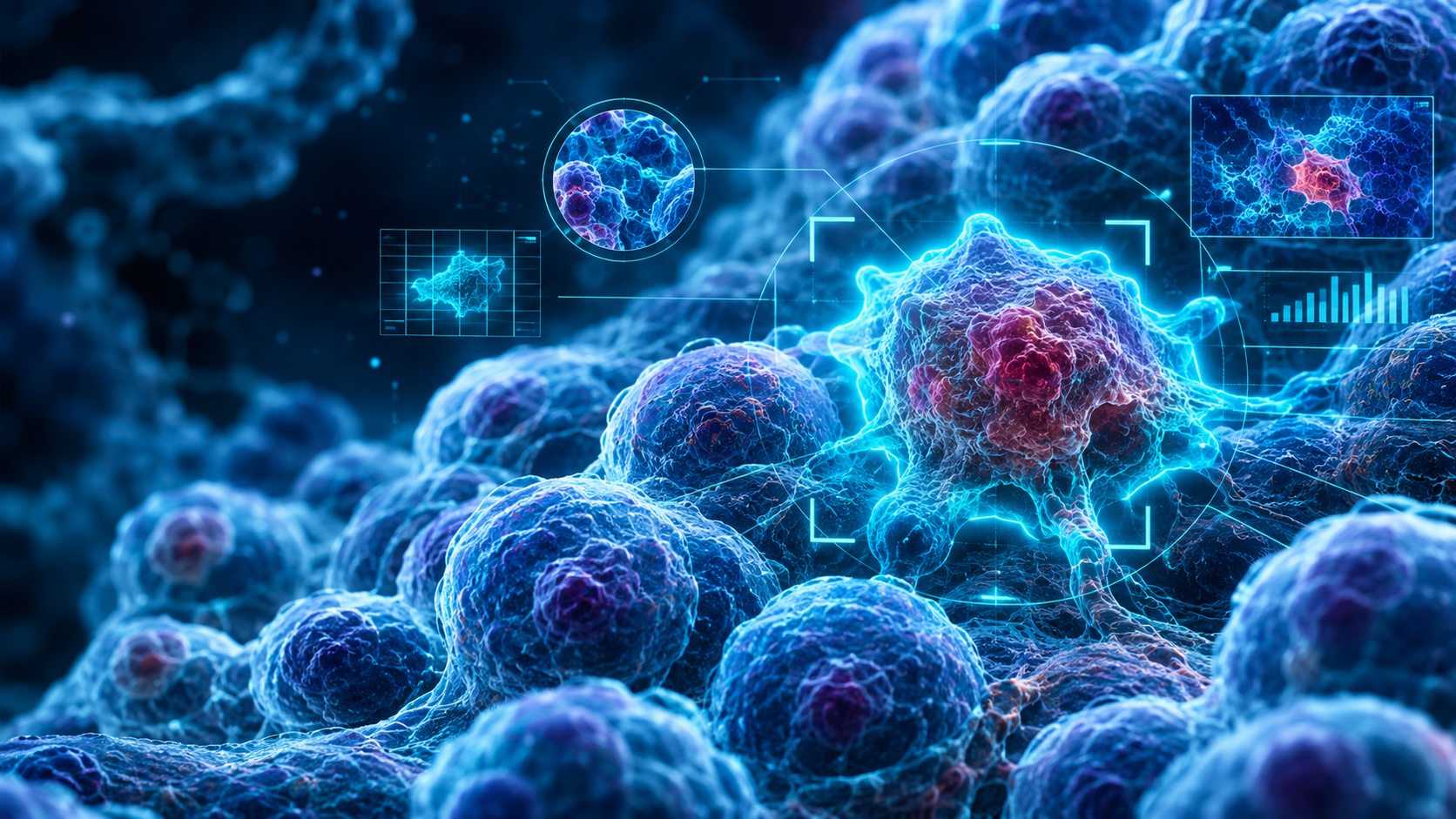
Cancer develops when cells acquire changes that allow them to grow and divide uncontrollably. As these cells multiply, they can disrupt normal tissue organization and form tumors.
Under a microscope, cancerous tissue may display several recognizable characteristics.
Abnormal Cell Morphology
Cell morphology refers to the shape, size, and structural appearance of cells.
Healthy cells generally maintain organized structures and relatively predictable patterns. Cancer cells, however, may display:
- Enlarged or irregular nuclei.
- Abnormal nuclear-to-cytoplasmic ratios.
- Variations in cell size and shape.
- Increased and abnormal mitotic activity.
- Irregular chromatin distribution.
These features, collectively associated with cellular atypia, can help pathologists distinguish malignant tissue from normal or benign tissue.
Changes in Tissue Architecture
Cancer does not only change individual cells. It can also alter how cells are organized within tissues.
Depending on the cancer type, pathologists may observe disrupted glandular structures, abnormal cell arrangements, invasive growth patterns, and loss of normal tissue organization.
These architectural changes can provide important evidence of malignancy.
The Tumor Microenvironment
A tumor is not made up exclusively of cancer cells.
It also contains blood vessels, fibroblasts, immune cells, connective tissue, and other components that collectively form the tumor microenvironment.
The interactions between these components can influence tumor growth, immune responses, invasion, and treatment resistance.
Understanding this environment is particularly important because two tumors with similar appearances may behave differently due to differences in their surrounding biological environments.
AI offers researchers a way to analyze these complex cellular relationships on a much larger scale.
2. How AI Examines Cancer Tissue: From Microscope Slides to Digital Intelligence
Traditional pathology involves placing thin tissue sections onto glass slides, staining them, and examining them under a microscope.
Digital pathology transforms this process by converting glass slides into high-resolution digital images.
These images are known as Whole Slide Images (WSIs).
A single whole-slide image can contain billions of pixels, making it far too large for most conventional image-processing systems to analyze as one complete image.
AI addresses this challenge through specialized image-processing techniques.
Step 1: Digitizing Tissue Samples
Before AI can analyze a tissue sample, it must be converted into a digital image.
A specialized whole-slide scanner captures microscopic images of the stained tissue, often using hematoxylin and eosin (H&E) staining.
H&E staining is widely used in pathology:
- Hematoxylin stains cell nuclei in shades of blue or purple.
- Eosin stains cytoplasm and extracellular structures in shades of pink.
These contrasting colors help reveal cellular organization and tissue abnormalities.
Once scanned, the image can be stored, examined, and processed using computational tools.
Step 2: Breaking Images Into Smaller Regions
Because whole-slide images are extremely large, AI systems commonly divide them into smaller regions called patches or tiles.
Each region contains a limited portion of the original tissue.
The algorithm analyzes these smaller sections individually before combining the extracted information to generate a broader interpretation of the entire slide.
This approach allows AI models to examine microscopic details while maintaining information about the overall tissue structure.
Step 3: Learning Cellular Patterns Through Deep Learning
Deep learning is a branch of machine learning that uses artificial neural networks to recognize complex patterns in data.
In computational pathology, convolutional neural networks (CNNs) have traditionally been among the most widely investigated architectures.
CNNs can learn visual features such as:
- Nuclear shape and arrangement.
- Cellular boundaries.
- Tissue density.
- Glandular structures.
- Tumor margins.
- Abnormal cellular patterns.
Rather than relying entirely on manually programmed rules, these networks learn patterns from large collections of annotated tissue images.
During training, the algorithm receives examples of tissue images alongside reference labels provided by pathologists. It gradually adjusts its internal parameters to recognize patterns associated with different tissue types and diagnostic categories.
Once trained, the model can analyze previously unseen images and generate predictions.
Step 4: Combining Microscopic Findings
Recognizing an abnormal cell in isolation does not necessarily establish a cancer diagnosis.
The surrounding tissue architecture is equally important.
Some AI systems therefore use a technique called Multiple Instance Learning (MIL).
In this approach, an algorithm examines multiple image patches from the same tissue sample and combines their information to make a slide-level prediction.
This is particularly useful when only a small region of a large tissue sample contains cancerous cells.
The system can identify areas that contribute most strongly to its prediction and direct the pathologist toward regions requiring closer examination.
3. What Can AI Actually Detect Inside Cancerous Tissue?
One of the most promising aspects of computational pathology is its ability to perform several different forms of tissue analysis.
Rather than simply identifying whether cancer is present, AI can assist with more detailed assessments.
A. Detecting Small Cancerous Regions
Some tissue samples contain only a limited number of malignant cells surrounded by large areas of normal tissue.
Finding these small regions can be difficult, particularly when the sample contains complex structures or abnormalities that resemble cancer.
AI models can scan large digital slides and highlight suspicious areas for further examination.
This may help identify small tumor deposits, abnormal glands, or metastatic cancer cells in lymph node tissue.
B. Identifying Tumor Boundaries
Understanding where a tumor begins and ends is important for determining its extent.
AI-based segmentation systems can distinguish tumor tissue from surrounding normal structures.
This process is useful in measuring tumor regions, estimating tumor cellularity, and evaluating the relationship between malignant tissue and surrounding structures.
However, distinguishing viable cancer cells from inflammation, fibrosis, necrosis, and treatment-related changes remains challenging.
C. Measuring Tumor Grade
Cancer grading involves assessing how abnormal tumor cells appear and how their structures differ from normal tissue.
For certain cancers, grading systems use specific microscopic features to estimate tumor aggressiveness.
AI can quantify these features and provide additional consistency during grading.
For example, in prostate cancer, computational models have been investigated for analyzing glandular architecture and supporting Gleason grading.
Such systems may assist pathologists in evaluating complex biopsy samples, although final interpretation must account for the relevant clinical and pathological criteria.
D. Analyzing Immune Cells Around Tumors
The immune system plays an important role in cancer development and treatment.
Some tumors contain large numbers of immune cells, while others develop environments that suppress immune activity.
AI can help quantify immune-cell populations, identify their spatial distribution, and measure how they are arranged in relation to cancer cells.
These measurements may provide useful information for investigating tumor immunity and understanding potential responses to immunotherapy.
E. Recognizing Microscopic Patterns Associated With Tumor Behavior
Researchers are also exploring whether AI can identify patterns associated with:
- Tumor aggressiveness.
- Lymph node metastasis.
- Recurrence risk.
- Molecular alterations.
- Treatment response.
- Patient prognosis.
Importantly, an image-based prediction is not automatically proof that a particular molecular alteration exists. These predictions require appropriate laboratory confirmation and clinical validation before they can guide treatment decisions.
4. Beyond What the Human Eye Can Measure: AI and Hidden Biological Patterns
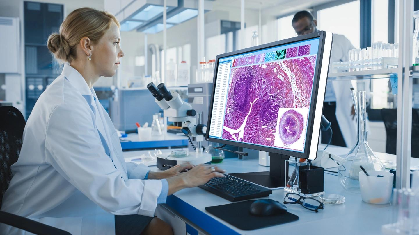
Perhaps the most interesting development in computational pathology is that AI may extract information from tissue images beyond traditional visual classification.
Pathologists are trained to recognize cellular abnormalities and tissue architecture. However, some biological patterns involve complex combinations of microscopic features that are difficult to describe or measure consistently.
AI can analyze thousands of visual characteristics simultaneously.
Computational Morphology
Computational morphology involves measuring the physical characteristics of cells and tissues using image-analysis algorithms.
These measurements may include nuclear size, shape irregularity, cellular density, spatial arrangement, and tissue organization.
By combining these features, researchers can investigate relationships between tissue appearance and cancer biology.
Predicting Molecular Information From Tissue Images
One particularly interesting research direction involves using AI to predict molecular characteristics from routine histology images.
In some studies, deep learning models have identified image patterns associated with gene mutations, molecular subtypes, or protein-expression patterns.
This is important because molecular testing can require additional laboratory procedures, specialized equipment, and resources.
However, image-based prediction should not be confused with direct genetic sequencing or definitive molecular testing.
AI may identify associations that help prioritize further investigation, but its predictions must be validated against appropriate molecular reference tests.
Foundation Models in Computational Pathology
Recent advances in AI have introduced pathology foundation models.
Unlike conventional models developed for one narrow diagnostic task, foundation models are trained on large collections of tissue images to learn more general visual representations.
These representations can potentially be adapted to different tasks, including tumor classification, tissue segmentation, biomarker prediction, and prognosis research.
Researchers are also exploring multimodal systems that combine pathology images with genomic information, clinical records, and other medical data.
This could eventually support more comprehensive approaches to understanding individual tumors.
5. Real-World Research: What Has Science Demonstrated?
The potential of AI in cancer pathology is supported by a growing body of scientific research.
However, it is important to distinguish promising experimental results from technologies that have already demonstrated reliable performance in routine clinical practice.
Research 1: Deep Learning in Histopathology
A major review published in Nature Medicine in 2021 examined the development of deep learning methods in histopathology.
The researchers described how AI systems had achieved performance comparable to trained pathologists in selected tasks, including tumor detection and grading.
However, the review also highlighted a major challenge: translating promising experimental algorithms into reliable clinical applications.
The authors emphasized the importance of validation, reproducibility, and integration into existing pathology workflows.
Research 2: AI Accuracy Across Digital Pathology Studies
A systematic review and meta-analysis published in npj Digital Medicine in 2024 evaluated AI-based diagnostic accuracy in digital pathology.
The analysis included 100 studies, with 48 contributing to the meta-analysis, and covered more than 152,000 whole-slide images.
The researchers reported:
- Mean sensitivity of 96.3%.
- Mean specificity of 93.3%.
Sensitivity describes how effectively a test identifies cases with the target condition, while specificity measures how effectively it identifies cases without that condition.
These results demonstrate the potential of AI in digital pathology.
However, an important limitation must be considered: 99% of the included studies had at least one area with a high or unclear risk of bias or applicability concerns.
Furthermore, the results covered different diseases and diagnostic tasks. They should not be interpreted as the accuracy of one universal AI system for detecting every type of cancer.
Research 3: AI and Personalized Cancer Treatment
A review published in the British Journal of Cancer in September 2025 explored how AI is transforming histological assessment.
It discussed applications extending beyond cancer detection, including:
- Tumor classification.
- Prognostic assessment.
- Prediction of genetic alterations.
- Potential treatment-response prediction.
- Personalized cancer management.
This research illustrates how computational pathology is gradually moving from simple image classification toward more comprehensive analysis of tumor biology.
6. Can AI Detect Cancer Better Than Experienced Pathologists?
The answer is more complicated than a simple yes or no.
AI can perform exceptionally well in carefully defined diagnostic tasks, particularly when analyzing large numbers of digital images.
It may also provide consistent quantitative measurements and identify areas that deserve additional attention.
However, cancer diagnosis requires much more than recognizing visual patterns.
A pathologist must consider the patient’s clinical history, tissue preparation, tumor location, previous treatments, laboratory findings, and the possibility of uncommon or misleading tissue appearances.
AI systems may perform poorly when they encounter samples that differ significantly from the data used during training.
For this reason, the most realistic approach is collaboration between AI systems and medical specialists.
Where AI Has an Advantage
- Rapid screening of large digital tissue collections.
- Consistent measurement of selected cellular features.
- Highlighting suspicious regions.
- Quantitative assessment of tumor characteristics.
- Supporting reproducibility in selected grading tasks.
Where Human Pathologists Remain Essential
- Interpreting unusual or rare pathological patterns.
- Integrating clinical information.
- Resolving ambiguous diagnostic findings.
- Evaluating unexpected or conflicting test results.
- Making final diagnostic judgments within the clinical context.
The future of computational pathology is therefore more likely to involve AI-assisted diagnosis than the complete replacement of pathologists.
7. The Major Challenges Preventing AI From Becoming a Universal Cancer Detection Tool
Despite significant progress, several scientific and technical barriers remain.
A. Differences Between Hospitals and Laboratories
Tissue samples can look different depending on how they are collected, preserved, stained, and scanned.
Even small variations in staining intensity, scanner settings, or tissue preparation can influence AI predictions.
A model trained using images from one hospital may not perform equally well in another institution.
This problem is commonly known as domain shift.
B. Limited and Biased Training Data
AI models depend heavily on the quality and diversity of their training datasets.
If a dataset contains mostly samples from a limited population, institution, or cancer subtype, the resulting model may not generalize to other groups.
Rare cancers and unusual tissue patterns are particularly difficult because relatively few annotated examples may be available.
C. The Black-Box Problem
Many deep learning models produce predictions without providing an easily understandable explanation of how they reached a conclusion.
In medicine, this creates a significant challenge.
Pathologists need to understand whether an algorithm is focusing on meaningful tumor characteristics or unrelated visual patterns.
Heatmaps and explainable AI techniques can help highlight important image regions, but they do not always reveal the actual biological reasoning behind a prediction.
D. Clinical Validation and Regulatory Approval
A model that performs well in a research experiment is not automatically suitable for patient care.
Before clinical implementation, AI systems need rigorous evaluation using appropriate datasets, independent testing, and real-world clinical workflows.
Regulatory requirements also depend on the intended use of the technology and the jurisdiction in which it is deployed.
The US Food and Drug Administration regulates relevant AI-enabled medical devices through established medical-device pathways.
E. Integration Into Existing Healthcare Systems
Hospitals need compatible scanners, digital storage infrastructure, secure data systems, trained staff, and reliable software integration.
These requirements can create financial and operational barriers, especially in healthcare systems with limited resources.
8. The Future of Cancer Diagnosis: Toward AI-Assisted Precision Pathology
The next generation of computational pathology may move beyond identifying cancerous cells toward understanding the biological behavior of entire tumors.
Several emerging research directions could shape this transformation.
Multimodal AI
Future systems may combine histopathology images with genomic sequencing, proteomic measurements, radiological scans, and clinical information.
Combining these different data sources could provide a more complete picture of tumor biology.
Spatial Analysis of Tumor Microenvironments
Spatial biology examines where different cells are located and how they interact within tissues.
AI could help researchers map the distribution of cancer cells, immune cells, and supporting tissue structures.
This may improve understanding of immune responses, tumor invasion, and treatment resistance.
More Personalized Cancer Treatment
If AI systems can reliably identify validated patterns associated with specific molecular characteristics or treatment outcomes, they may help clinicians select patients for additional testing or appropriate therapies.
However, treatment decisions will continue to require validated biomarkers, clinical evidence, and specialist judgment.
Faster and More Consistent Pathology Workflows
As digital pathology becomes more widely adopted, AI may help automate repetitive image-analysis tasks, prioritize suspicious cases, and reduce the time required for certain assessments.
The greatest benefit may come from allowing pathologists to concentrate more attention on difficult and complex cases.
9. Can AI Eventually See What Humans Cannot?
The most important development in AI-powered pathology may not be that machines can recognize cancer faster than humans.
It may be their ability to measure complex patterns that are difficult for humans to quantify consistently.
A tissue sample contains enormous amounts of biological information. The arrangement of cells, the structure of their nuclei, the organization of surrounding tissue, and the spatial distribution of immune cells can all provide clues about tumor behavior.
AI offers a way to examine these patterns at a scale that conventional manual analysis cannot easily reproduce.
Yet, recognizing a pattern is not the same as understanding the disease. A prediction is not automatically a diagnosis, and a statistical association does not establish a biological mechanism.
The future of cancer pathology will depend on combining computational power with biological knowledge, clinical experience, and rigorous scientific validation.
AI may not replace the pathologist examining the tissue. Instead, it could give that pathologist an entirely new way to see it.
Conclusion
Artificial intelligence is changing how researchers and medical specialists examine cancer at the microscopic level.
Through digital pathology, deep learning, cellular image analysis, and multimodal data integration, AI systems can identify complex tissue patterns, quantify tumor characteristics, and support investigations into cancer progression and treatment response.
Although many applications remain under evaluation, the scientific progress is significant.
The transition from experimental algorithms to dependable clinical tools will require larger and more diverse datasets, independent validation, transparent performance reporting, and appropriate regulatory oversight.
Ultimately, the future of cancer diagnosis may not belong to humans or machines independently. It may emerge from their ability to work together, combining the interpretive expertise of pathologists with the analytical capabilities of artificial intelligence.
References and Further Reading
- van der Laak, J., Litjens, G., & Ciompi, F. (2021). Deep learning in histopathology: the path to the clinic. Nature Medicine.
https://doi.org/10.1038/s41591-021-01343-4 - McGenity, C. et al. (2024). Artificial intelligence in digital pathology: a systematic review and meta-analysis of diagnostic test accuracy. npj Digital Medicine.
https://doi.org/10.1038/s41746-024-01106-8 - Takamatsu, M. (2025). Transforming histologic assessment: artificial intelligence in cancer diagnosis and personalized treatment. British Journal of Cancer.
https://doi.org/10.1038/s41416-025-03206-y - Baxi, V. et al. (2022). Digital pathology and artificial intelligence in translational medicine and clinical practice. Modern Pathology.
https://doi.org/10.1038/s41379-021-00919-2 - Artificial intelligence and digital pathology: clinical promise and deployment considerations (2023). Journal of Medical Imaging.
https://pmc.ncbi.nlm.nih.gov/articles/PMC10389766/ - US Food and Drug Administration. Artificial Intelligence-Enabled Medical Devices.
https://www.fda.gov/medical-devices/digital-health-center-excellence/artificial-intelligence-enabled-medical-devices - Translational AI in whole-slide image cancer histopathology: state of the art and regulatory-approved solutions (2026). Frontiers in Digital Health.
https://doi.org/10.3389/fdgth.2026.1863382